Automated Muscle and Fat Segmentation in Computed Tomography for Comprehensive Body Composition Analysis
Yaqian Chen1, Hanxue Gu1, Yuwen Chen1, Jicheng Yang1, Haoyu Dong1, Joseph Y. Cao2, Adrian Camarena3, Christopher Mantyh3, Roy Colglazier2, Maciej A. Mazurowski4,2,5,6
1: Department of Electrical and Computer Engineering, Duke University, Durham, NC 27708, 2: Department of Radiology, Duke University, Durham, NC 27708, 3: Department of Surgery Duke University School of Medicine, Durham, NC 27708 , 4: Department of Electrical and Computer Engineering, Duke University, Durham, NC 27708 , 5: Department of Biostatistics & Bioinformatics, Duke University, Durham, NC 27708 , 6: Department of Computer Science, Duke University, Durham, NC 27708
Publication date: 2025/11/16
https://doi.org/10.59275/j.melba.2025-ag5g
Abstract
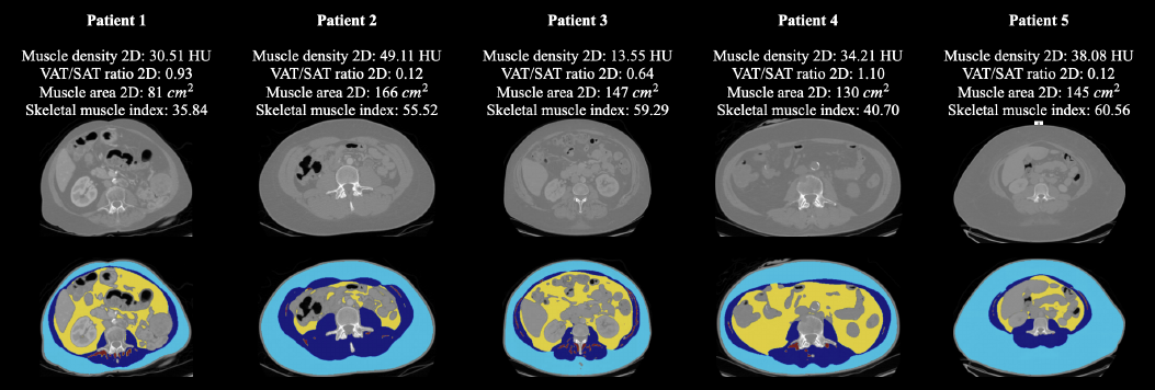
Body composition assessment using CT images can potentially be used for a number of clinical applications, including the prognostication of cardiovascular outcomes, evaluation of metabolic health, monitoring of disease progression, assessment of nutritional status, prediction of treatment response in oncology, and risk stratification for surgical and critical care outcomes. While multiple groups have developed in-house segmentation tools for this analysis, there are very limited publicly available tools that could be consistently used across di!erent applications. To mitigate this gap, we present a publicly accessible, end-to-end segmentation and feature calculation model specifically for CT body composition analysis. Our model performs segmentation of skeletal muscle, subcutaneous adipose tissue (SAT), and visceral adipose tissue (VAT) across the chest, abdomen, and pelvis area in axial CT images. It also provides various body composition metrics, including muscle density, visceral-to-subcutaneous fat (VAT/SAT) ratio, muscle area/volume, and skeletal muscle index (SMI), supporting both 2D and 3D assessments. To evaluate the model, the segmentation was applied to both internal and external datasets, with body composition metrics analyzed across di!erent age, sex, and race groups. The model achieved high dice coe”cients on both internal and external datasets, exceeding 89% for skeletal muscle, SAT, and VAT segmentation. The model outperforms the benchmark by 2.40% on skeletal muscle and 10.26% on SAT compared to the manual annotations given by the publicly available dataset. Body composition metrics show mean relative absolute errors (MRAEs) under 10% for all measures. Furthermore, the model provided muscular fat segmentation with a Dice coe”cient of 56.27%, which can be utilized for additional analyses as needed. Our model with weights is publicly available at https://github.com/mazurowski-lab/CT-Muscle-and-Fat-Segmentation.git
Keywords
Deep learning · Segmentation · Muscles · Subcutaneous Fat · Visceral Fat · Body Composition
Bibtex
@article{melba:2025:026:chen,
    title = "Automated Muscle and Fat Segmentation in Computed Tomography for Comprehensive Body Composition Analysis",
    author = "Chen, Yaqian and Gu, Hanxue and Chen, Yuwen and Yang, Jicheng and Dong, Haoyu and Cao, Joseph Y. and Camarena, Adrian and Mantyh, Christopher and Colglazier, Roy and Mazurowski, Maciej A.",
    journal = "Machine Learning for Biomedical Imaging",
    volume = "3",
    issue = "November 2025 issue",
    year = "2025",
    pages = "581--618",
    issn = "2766-905X",
    doi = "https://doi.org/10.59275/j.melba.2025-ag5g",
    url = "https://melba-journal.org/2025:026"
}
RIS
TY - JOUR
AU - Chen, Yaqian
AU - Gu, Hanxue
AU - Chen, Yuwen
AU - Yang, Jicheng
AU - Dong, Haoyu
AU - Cao, Joseph Y.
AU - Camarena, Adrian
AU - Mantyh, Christopher
AU - Colglazier, Roy
AU - Mazurowski, Maciej A.
PY - 2025
TI - Automated Muscle and Fat Segmentation in Computed Tomography for Comprehensive Body Composition Analysis
T2 - Machine Learning for Biomedical Imaging
VL - 3
IS - November 2025 issue
SP - 581
EP - 618
SN - 2766-905X
DO - https://doi.org/10.59275/j.melba.2025-ag5g
UR - https://melba-journal.org/2025:026
ER -