MyData: A Comprehensive Database of Mycetoma Tissue Microscopic Images for Histopathological Analysis
Hyam Omar Ali1,2,3,4, Romain Abraham4, Guillaume Desoubeaux5,6, Ahmed Fahal2, Clovis Tauber3
1: Faculty of Mathematical Sciences, University of Khartoum, 11111, Khartoum, Sudan, 2: The Mycetoma Research Centre, University of Khartoum, 11111, Khartoum, Sudan, 3: U1253 iBrain, University of Tours, Inserm, 37032, Tours, France, 4: CNRS U7013 Institute Denis Poisson, University of Orleans, 45067, Orleans, France, 5: Parasitology and Mycology Department, Bretonneau Hospital, 37032, Tours, France, 6: INSERM U1100, University of Tours, 37032, Tours, France
Publication date: 2025/12/31
https://doi.org/10.59275/j.melba.2025-5791
Abstract
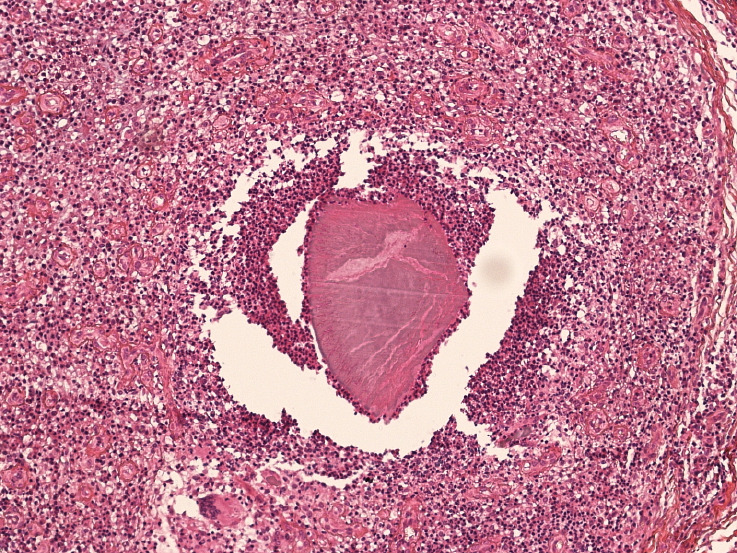
Mycetoma is a chronic and neglected inflammatory disease prevalent in tropical and subtropical regions. It can lead to severe disability and social stigma. The disease is classified into two types based on the causative microorganisms: eumycetoma (fungal) and actinomycetoma (bacterial). Effective treatment strategies depend on accurately identifying the causative agents. Current identification methods include molecular, cytological, and histopathological techniques, as well as grain culturing. Among these, histopathological techniques are considered optimal for use in endemic areas, but they require expert pathologists for accurate identification, which can be challenging in rural areas lacking such expertise. The advent of digital pathology and automated image analysis algorithms offers a potential solution. This report introduces a novel dataset designed for the automated detection and classification of mycetoma using histopathological images. It includes the first database of microscopic images of mycetoma tissue, detailing the entire pipeline from species distribution and patient sampling to acquisition protocols through histological procedures. The dataset consists of images from 142 patients, totalling 864 images, each annotated with binary masks indicating the presence of grains, facilitating both detection and segmentation tasks.
Keywords
Mycetoma · histopathology diagnosis · Microscopic images · Image Analysis · Classification · Segmentation
Bibtex
@article{melba:2025:045:omarali,
    title = "MyData: A Comprehensive Database of Mycetoma Tissue Microscopic Images for Histopathological Analysis",
    author = "Omar Ali, Hyam and Abraham, Romain and Desoubeaux, Guillaume and Fahal, Ahmed and Tauber, Clovis",
    journal = "Machine Learning for Biomedical Imaging",
    volume = "3",
    issue = "Special Issue on MICCAI Open Data 2024-2025",
    year = "2025",
    pages = "897--904",
    issn = "2766-905X",
    doi = "https://doi.org/10.59275/j.melba.2025-5791",
    url = "https://melba-journal.org/2025:045"
}
RIS
TY - JOUR
AU - Omar Ali, Hyam
AU - Abraham, Romain
AU - Desoubeaux, Guillaume
AU - Fahal, Ahmed
AU - Tauber, Clovis
PY - 2025
TI - MyData: A Comprehensive Database of Mycetoma Tissue Microscopic Images for Histopathological Analysis
T2 - Machine Learning for Biomedical Imaging
VL - 3
IS - Special Issue on MICCAI Open Data 2024-2025
SP - 897
EP - 904
SN - 2766-905X
DO - https://doi.org/10.59275/j.melba.2025-5791
UR - https://melba-journal.org/2025:045
ER -