Benchmarking Foundation Models for Mitotic Figure Classification
Jonas Ammeling1 , Jonathan Ganz2,1, Emely Rosbach1, Ludwig Lausser1, Christof A. Bertram3, Katharina Breininger4,5, Marc Aubreville6
1: Technische Hochschule Ingolstadt, AImotion, Ingolstadt, DE, 2: MIRA vision Microscopy GmbH, Göppingen, DE, 3: University of Veterinary Medicine, Institute of Pathology, Vienna, AU, 4: Center for AI and Data Science, Julius-Maximilians-Universität Würzburg, Würzburg, DE, 5: Department Artificial Intelligence in Biomedical Engineering, Friedrich-Alexander-Universität Erlangen-Nürnberg, Erlangen, DE, 6: Flensburg University of Applied Sciences, Flensburg, DE
Publication date: 2026/01/30
https://doi.org/10.59275/j.melba.2026-a3eb
Abstract
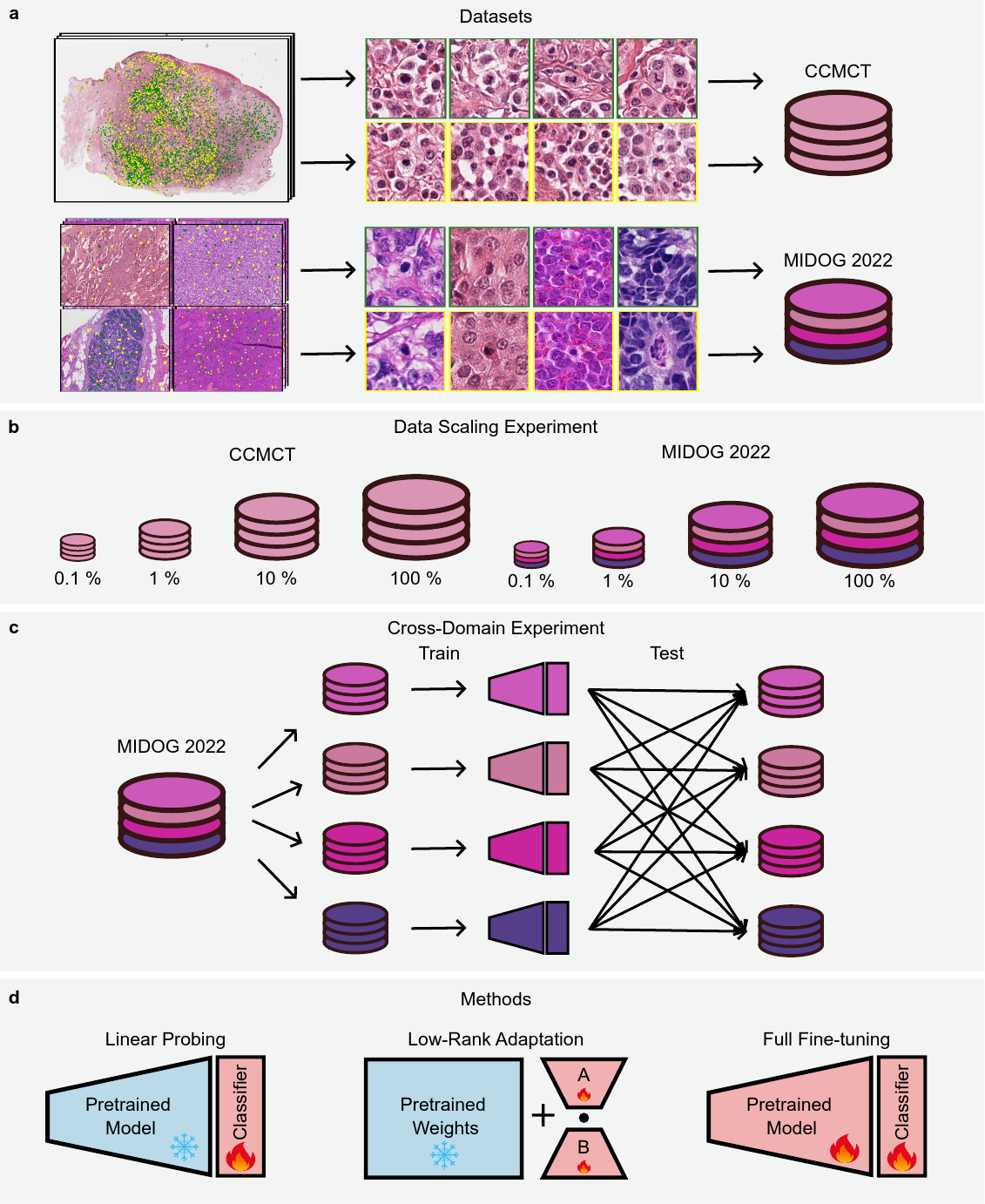
The performance of deep learning models is known to scale with data quantity and diversity. In pathology, as in many other medical imaging domains, the availability of labeled images for a specific task is often limited. Self-supervised learning techniques have enabled the use of vast amounts of unlabeled data to train large-scale neural networks, i.e., foundation models, that can address the limited data problem by providing semantically rich feature vectors that can generalize well to new tasks with minimal training effort increasing model performance and robustness. In this work, we investigate the use of foundation models for mitotic figure classification. The mitotic count, which can be derived from this classification task, is an independent prognostic marker for specific tumors and part of certain tumor grading systems. In particular, we investigate the data scaling laws on multiple current foundation models and evaluate their robustness to unseen tumor domains. Next to the commonly used linear probing paradigm, we also adapt the models using low-rank adaptation (LoRA) of their attention mechanisms. We compare all models against end-to-end-trained baselines, both CNNs and Vision Transformers. Our results demonstrate that LoRA-adapted foundation models provide superior performance to those adapted with standard linear probing, reaching performance levels close to 100 % data availability with only 10 % of training data. Furthermore, LoRA-adaptation of the most recent foundation models almost closes the out-of-domain performance gap when evaluated on unseen tumor domains. However, full fine-tuning of traditional architectures still yields competitive performance.
Keywords
Machine Learning · Computational Pathology · Self-Supervised Learning · Foundation Models · Mitotic Figure Classification · Low-Rank Adaptation
Bibtex
@article{melba:2026:003:ammeling,
    title = "Benchmarking Foundation Models for Mitotic Figure Classification",
    author = "Ammeling, Jonas and Ganz, Jonathan and Rosbach, Emely and Lausser, Ludwig and Bertram, Christof A. and Breininger, Katharina and Aubreville, Marc",
    journal = "Machine Learning for Biomedical Imaging",
    volume = "3",
    issue = "MELBA–BVM 2025 Special Issue",
    year = "2026",
    pages = "38--58",
    issn = "2766-905X",
    doi = "https://doi.org/10.59275/j.melba.2026-a3eb",
    url = "https://melba-journal.org/2026:003"
}
RIS
TY - JOUR
AU - Ammeling, Jonas
AU - Ganz, Jonathan
AU - Rosbach, Emely
AU - Lausser, Ludwig
AU - Bertram, Christof A.
AU - Breininger, Katharina
AU - Aubreville, Marc
PY - 2026
TI - Benchmarking Foundation Models for Mitotic Figure Classification
T2 - Machine Learning for Biomedical Imaging
VL - 3
IS - MELBA–BVM 2025 Special Issue
SP - 38
EP - 58
SN - 2766-905X
DO - https://doi.org/10.59275/j.melba.2026-a3eb
UR - https://melba-journal.org/2026:003
ER -