Robust Renal Mass Segmentation on CT: A Validation Study of an AI-Based Framework
Sarah de Boer1 , Hartmut Häntze1,2, Kiran Vaidhya Venkadesh1, Myrthe A. D. Buser1, Gabriel E. Humpire Mamani1, Lina Xu2, Lisa C. Adams3, Jawed Nawabi4, Keno K. Bressem3,5, Bram van Ginneken1,6, Mathias Prokop1, Alessa Hering1
1: Department of Medical Imaging, Radboudumc, Nijmegen, The Netherlands, 2: Department of Radiology, Charité - Universitätsmedizin Berlin, Berlin, Germany, 3: Department of Diagnostic and Interventional Radiology, Klinikum rechts der Isar, TUM University Hospital, Technical University of Munich, Munich, Germany, 4: Department of Neuroradiology, Charité - Universitätsmedizin Berlin, Berlin, Germany, 5: Department of Cardiovascular Radiology and Nuclear Medicine, German Heart Center, TUM University Hospital, Technical University of Munich, Munich, Germany, 6: Fraunhofer MEVIS, Bremen, Germany
Publication date: 2026/05/22
https://doi.org/10.59275/j.melba.2026-67g5
Abstract
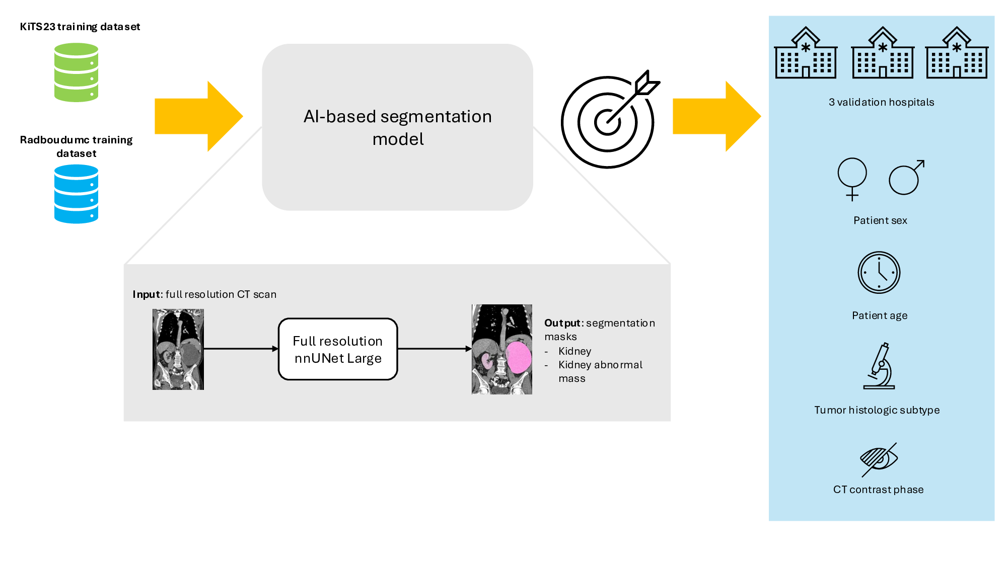
Renal mass segmentation has important potential to enhance the clinical workflow, especially in settings requiring quantitative assessments. Kidney volume could serve as an important biomarker for renal diseases, with changes in volume correlating directly with kidney function. Currently, clinical practice often relies on subjective visual assessment for evaluating kidney size and kidney lesions, including tumors and cysts, which are typically staged based on diameter, volume, and anatomical location. To support a more objective and reproducible approach, this research aims to develop a robust, thoroughly validated renal mass segmentation algorithm, named Renal-Net. We employ publicly available training datasets and leverage the state-of-the-art medical image segmentation framework nnU-Net. Validation is conducted using both proprietary and public test datasets, with segmentation performance quantified by Dice coefficient and the 95th percentile Hausdorff distance. Furthermore, we analyze robustness across subgroups based on patient sex, age, CT contrast phases, and tumor histologic subtypes. Our findings demonstrate that our segmentation algorithm, trained exclusively on publicly available data, generalizes effectively to external test sets and outperforms existing state-of-the-art models across all tested datasets. Subgroup analyses reveal consistent high performance, indicating strong robustness and reliability. The developed algorithm and associated code are publicly accessible at https://github.com/DIAGNijmegen/oncology-kidney-abnormality-segmentation
Keywords
Deep Learning · Medical Imaging · Segmentation · Kidney cancer · Renal Cell Carcinoma · Renal mass
Bibtex
@article{melba:2026:012:deboer,
    title = "Robust Renal Mass Segmentation on CT: A Validation Study of an AI-Based Framework",
    author = "de Boer, Sarah and Häntze, Hartmut and Venkadesh, Kiran Vaidhya and Buser, Myrthe A. D. and Humpire Mamani, Gabriel E. and Xu, Lina and Adams, Lisa C. and Nawabi, Jawed and Bressem, Keno K. and van Ginneken, Bram and Prokop, Mathias and Hering, Alessa",
    journal = "Machine Learning for Biomedical Imaging",
    volume = "2026",
    issue = "May 2026 issue",
    year = "2026",
    pages = "229--251",
    issn = "2766-905X",
    doi = "https://doi.org/10.59275/j.melba.2026-67g5",
    url = "https://melba-journal.org/2026:012"
}
RIS
TY - JOUR
AU - de Boer, Sarah
AU - Häntze, Hartmut
AU - Venkadesh, Kiran Vaidhya
AU - Buser, Myrthe A. D.
AU - Humpire Mamani, Gabriel E.
AU - Xu, Lina
AU - Adams, Lisa C.
AU - Nawabi, Jawed
AU - Bressem, Keno K.
AU - van Ginneken, Bram
AU - Prokop, Mathias
AU - Hering, Alessa
PY - 2026
TI - Robust Renal Mass Segmentation on CT: A Validation Study of an AI-Based Framework
T2 - Machine Learning for Biomedical Imaging
VL - 2026
IS - May 2026 issue
SP - 229
EP - 251
SN - 2766-905X
DO - https://doi.org/10.59275/j.melba.2026-67g5
UR - https://melba-journal.org/2026:012
ER -