Structured Spectral Graph Representation Learning for Multi-label Abnormality Analysis from 3D CT Scans
Theo Di Piazza1,2, Carole Lazarus3, Olivier Nempont3, Loic Boussel1,2
1: INSA Lyon, University of Lyon, CNRS, INSERM, CREATIS UMR 5220, U1294, Villeurbanne, France, 2: Hospices Civil de Lyon, Lyon, France, 3: Philips Clinical Informatics, Innovation Paris, France
Publication date: 2026/06/15
https://doi.org/10.59275/j.melba.2026-87e3
Abstract
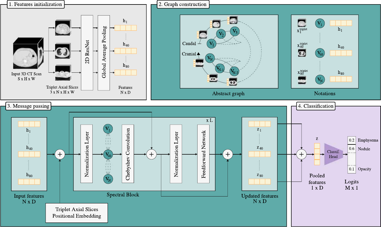
With the growing volume of CT examinations, there is an increasing demand for automated tools such as organ segmentation, abnormality detection, and report generation to support radiologists in managing their clinical workload. Multi-label classification of 3D Chest CT scans remains a critical yet challenging problem due to the complex spatial relationships inherent in volumetric data and the wide variability of abnormalities. Existing methods based on 3D convolutional neural networks struggle to capture long-range dependencies, while Vision Transformers often require extensive pre-training on large-scale, domain-specific datasets to perform competitively. In this work, we propose a 2.5D alternative by introducing a new graph-based framework that represents 3D CT volumes as structured graphs, where axial slice triplets serve as nodes processed through spectral graph convolution, enabling the model to reason over inter-slice dependencies while maintaining complexity compatible with clinical deployment. Our method, trained and evaluated on 3 datasets from independent institutions, achieves strong cross-dataset generalization, and shows competitive performance compared to state-of-the-art visual encoders. We further conduct comprehensive ablation studies to evaluate the impact of various aggregation strategies, edge-weighting schemes, and graph connectivity patterns. Additionally, we demonstrate the broader applicability of our approach through transfer experiments on automated radiology report generation and abdominal CT data. This work extends our previous contribution presented at the MICCAI 2025 EMERGE Workshop. A project page is available at https://theodpzz.github.io/projects/ctssg
Keywords
3D Medical Imaging · Computed Tomography · Representation Learning · Graph Neural Network · Spectral domain · Multi-label Abnormality Classification · Automated Report Generation
Bibtex
@article{melba:2026:018:dipiazza,
    title = "Structured Spectral Graph Representation Learning for Multi-label Abnormality Analysis from 3D CT Scans",
    author = "Di Piazza, Theo and Lazarus, Carole and Nempont, Olivier and Boussel, Loic",
    journal = "Machine Learning for Biomedical Imaging",
    volume = "2026",
    issue = "June 2026 issue",
    year = "2026",
    pages = "359--387",
    issn = "2766-905X",
    doi = "https://doi.org/10.59275/j.melba.2026-87e3",
    url = "https://melba-journal.org/2026:018"
}
RIS
TY - JOUR
AU - Di Piazza, Theo
AU - Lazarus, Carole
AU - Nempont, Olivier
AU - Boussel, Loic
PY - 2026
TI - Structured Spectral Graph Representation Learning for Multi-label Abnormality Analysis from 3D CT Scans
T2 - Machine Learning for Biomedical Imaging
VL - 2026
IS - June 2026 issue
SP - 359
EP - 387
SN - 2766-905X
DO - https://doi.org/10.59275/j.melba.2026-87e3
UR - https://melba-journal.org/2026:018
ER -