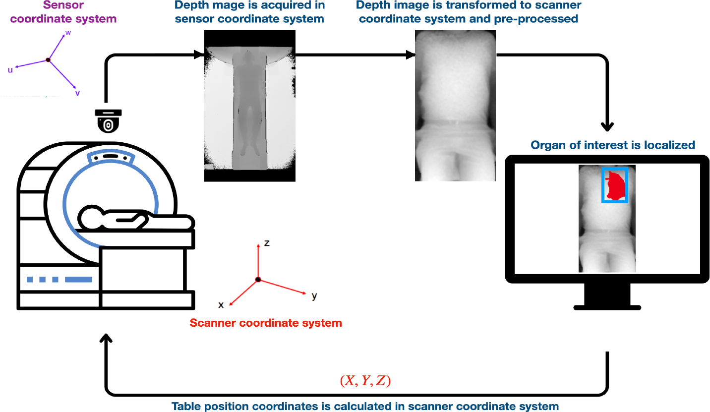
Depth to Anatomy: Organ Localization from Depth Images for Automated Patient Table Positioning in Radiology Workflow
Eytan Kats1, Kai Geissler2, Jochen G. Hirsch2, Daniel Mensing2, Julien Senegas3, Stefan Heldman2, Mattias P. Heinrich1
1: Institute of Medical Informatics, University of Luebeck, Luebeck, Germany, 2: Fraunhofer Institute for Digital Medicine MEVIS, Bremen, Germany, 3: Philips Research, Hamburg, Germany
Publication date: 2026/07/22
https://doi.org/10.59275/j.melba.2026-gc51
Abstract
Automated patient positioning can improve radiology workflow efficiency by reducing the time required for manual table adjustments and scout-based scan planning. We propose a learning-based framework that predicts 3D organ locations and shapes for 41 anatomical structures, including both bones and soft tissues, directly from a single 2D depth image of the body surface. Leveraging 10, 020 whole-body MRI scans from the German National Cohort (NAKO) dataset, we synthetically generate depth images paired with anatomical segmentations to train a convolutional neural network for volumetric organ prediction. Our method achieves a mean dice similarity coefficient of 0.44 ± 0.2 and and a symmetric average surface distance of 7.69 ± 5.68 mm across all structures. Furthermore, the model derives organ bounding boxes with a mean absolute detection offset of 10.99 ± 5.54 mm. Qualitative results on real-world depth images confirm the ability of the model to generalize to practical clinical settings. These findings suggest that depth-only organ localization can support automated patient positioning reducing setup time, minimizing operator variability, and improving patient comfort. The implementation and pretrained models are publicly available at https://github.com/EytanKats/orgloc
Keywords
Radiology workflow automation · Automated patient table positioning · Body surface-to-3D anatomy prediction · Hybrid 2D–to-3D neural architecture
Bibtex
@article{melba:2026:024:kats,
    title = "Depth to Anatomy: Organ Localization from Depth Images for Automated Patient Table Positioning in Radiology Workflow",
    author = "Kats, Eytan and Geissler, Kai and Hirsch, Jochen G. and Mensing, Daniel and Senegas, Julien and Heldman, Stefan and Heinrich, Mattias P.",
    journal = "Machine Learning for Biomedical Imaging",
    volume = "2026",
    issue = "MELBA–BVM 2025 Special Issue",
    year = "2026",
    pages = "507--522",
    issn = "2766-905X",
    doi = "https://doi.org/10.59275/j.melba.2026-gc51",
    url = "https://melba-journal.org/2026:024"
}
RIS
TY - JOUR
AU - Kats, Eytan
AU - Geissler, Kai
AU - Hirsch, Jochen G.
AU - Mensing, Daniel
AU - Senegas, Julien
AU - Heldman, Stefan
AU - Heinrich, Mattias P.
PY - 2026
TI - Depth to Anatomy: Organ Localization from Depth Images for Automated Patient Table Positioning in Radiology Workflow
T2 - Machine Learning for Biomedical Imaging
VL - 2026
IS - MELBA–BVM 2025 Special Issue
SP - 507
EP - 522
SN - 2766-905X
DO - https://doi.org/10.59275/j.melba.2026-gc51
UR - https://melba-journal.org/2026:024
ER -