The MYOSAIQ Challenge: Myocardial Segmentation with Automated Infarct Quantification
Olivier Bernard1,2, William A. Romero R.1, Cyprien Bouton1, Celia Goujat1, Hang Jung Ling1, Pierre-Marc Jodoin3, Fumin Guo4, Calder Sheagren5, Graham Wright5, Abdul Qayyum6, Moona Mazher7, Steven A. Niederer6, Hairui Wang7, Xiaomei Wu8, Franz Thaler9,10, Gernot Plank9, Martin Urschler11, Ricardo M. Rosales12, Esther Pueyo12, Nicolas Duchateau1,2, Frederic Cervenansky1, Patrick Clarysse1, Loic Belle13, Thomas Bochaton14,15, Nathan Mewton14,15, Magalie Viallon1,16, Pierre Croisille1,16
1: INSA-Lyon, Universite Claude Bernard Lyon 1, UJM-Saint Etienne, CNRS, Inserm, CREATIS UMR 5220, U1294, F-42023, Saint Etienne, France, 2: Institut Universitaire de France (IUF), France, 3: Department of Computer Science, University of Sherbrooke, Sherbrooke, QC, Canada, 4: Wuhan National Library of Optoelectronics, Huazhong University of Science and Technology, Wuhan, China, 5: Sunnybrook Research Institute, University of Toronto, Toronto, Canada, 6: National Heart and Lung Institute, Faculty of Medicine, Imperial College London, London, United Kingdom, 7: Centre for Medical Image Computing, Department of Computer Science, University College London, London, United Kingdom, 8: Fudan University, Shanghai, 200433, China, 9: Gottfried Schatz Research Center: Medical Physics and Biophysics, Medical University of Graz, Graz, Austria, 10: Institute of Computer Graphics and Vision, Graz University of Technology, Graz, Austria, 11: Institute for Medical Informatics, Statistics and Documentation, Medical University of Graz, Graz, Austria, 12: Instituto de Investigación en Ingenierı́a de Aragón, Universidad de Zaragoza; Instituto de Investigación Sanitaria de Aragón (IIS Aragón); CIBER-BBN, Zaragoza, Aragón, Spain, 13: Centre Hospitalier Annecy Genevois, France, 14: Hôpital Universitaire Cardiologique Louis Pradel, Bron, France, 15: CarMeN INSERM U1060, Universite Claude Bernard Lyon 1, Lyon, France, 16: Hôpital Universitaire de Saint-Etienne, Saint-Etienne, France
Publication date: 2026/08/28
https://doi.org/10.59275/j.melba.2026-e93b
Abstract
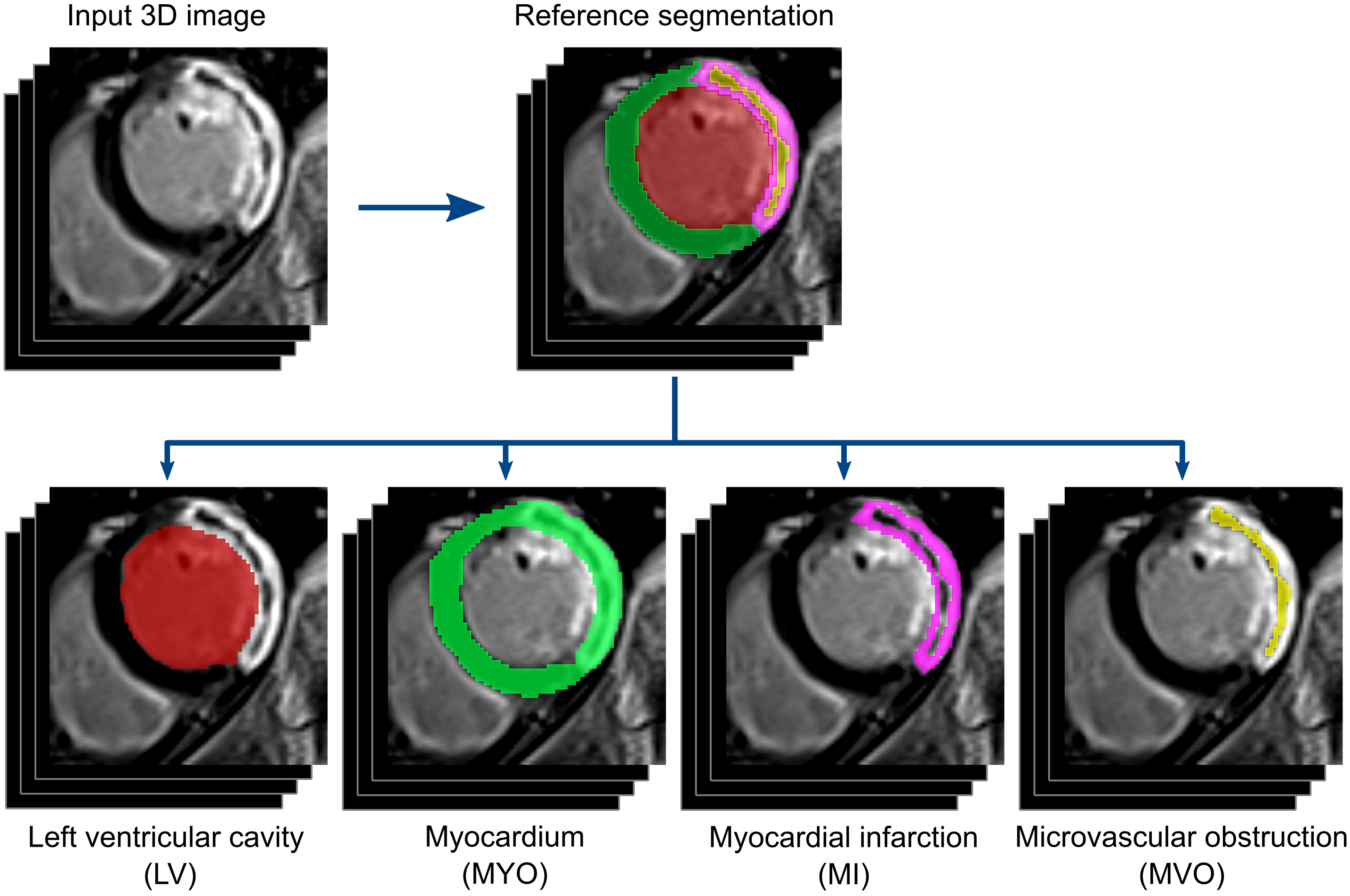
Late gadolinium enhancement (LGE) cardiac magnetic resonance (MR) imaging is the modality of choice to assess myocardial infarction (MI) lesions. Nowadays MI volume quantification is not performed routinely in clinical practice. Numerous deep learning (DL) methods have been developed to automate the segmentation of the myocardium and infarct regions. However, most studies rely on relatively small datasets which typically undergo pre-processing steps to standardize images and focus on a specific phase of myocardial infarction following reperfusion therapy. These limitations have impeded the development of models that are generalizable across diverse conditions and thus suitable for routine clinical use. To advance research and establish benchmarks in generalizable learning for myocardial infarct quantification, this paper presents findings from the Myocardial Segmentation with Automated Infarct Quantification (MYOSAIQ) challenge. The dataset set up for the challenge combines 439 CMR volumes from two multicenter clinical trials, with representative data acquired in acute and chronic phases after acute MI. Data were acquired in 16 centers using MRI scanners from three different vendors. Six teams participated until the end of the challenge, employing various baseline models, data augmentation techniques, and confidence strategies. To enhance the significance of this study, we compare the challengers’ results with those of fine-tuned foundation models. Our results indicate that well-designed UNet-based techniques outperform fully automatic foundation models for LGE MR segmentation. While the best methods achieve high-quality and stable delineations of the left ventricle and myocardium under various conditions, they remain improvable in accurately segmenting infarct regions.
Keywords
Cardiac imaging · Late Gadolinium Enhancement · segmentation · myocardial infarct
Bibtex
@article{melba:2026:032:bernard,
    title = "The MYOSAIQ Challenge: Myocardial Segmentation with Automated Infarct Quantification",
    author = "Bernard, Olivier and Romero R., William A. and Bouton, Cyprien and Goujat, Celia and Ling, Hang Jung and Jodoin, Pierre-Marc and Guo, Fumin and Sheagren, Calder and Wright, Graham and Qayyum, Abdul and Mazher, Moona and Niederer, Steven A. and Wang, Hairui and Wu, Xiaomei and Thaler, Franz and Plank, Gernot and Urschler, Martin and Rosales, Ricardo M. and Pueyo, Esther and Duchateau, Nicolas and Cervenansky, Frederic and Clarysse, Patrick and Belle, Loic and Bochaton, Thomas and Mewton, Nathan and Viallon, Magalie and Croisille, Pierre",
    journal = "Machine Learning for Biomedical Imaging",
    volume = "2026",
    issue = "August 2026 issue",
    year = "2026",
    pages = "658--673",
    issn = "2766-905X",
    doi = "https://doi.org/10.59275/j.melba.2026-e93b",
    url = "https://melba-journal.org/2026:032"
}
RIS
TY - JOUR
AU - Bernard, Olivier
AU - Romero R., William A.
AU - Bouton, Cyprien
AU - Goujat, Celia
AU - Ling, Hang Jung
AU - Jodoin, Pierre-Marc
AU - Guo, Fumin
AU - Sheagren, Calder
AU - Wright, Graham
AU - Qayyum, Abdul
AU - Mazher, Moona
AU - Niederer, Steven A.
AU - Wang, Hairui
AU - Wu, Xiaomei
AU - Thaler, Franz
AU - Plank, Gernot
AU - Urschler, Martin
AU - Rosales, Ricardo M.
AU - Pueyo, Esther
AU - Duchateau, Nicolas
AU - Cervenansky, Frederic
AU - Clarysse, Patrick
AU - Belle, Loic
AU - Bochaton, Thomas
AU - Mewton, Nathan
AU - Viallon, Magalie
AU - Croisille, Pierre
PY - 2026
TI - The MYOSAIQ Challenge: Myocardial Segmentation with Automated Infarct Quantification
T2 - Machine Learning for Biomedical Imaging
VL - 2026
IS - August 2026 issue
SP - 658
EP - 673
SN - 2766-905X
DO - https://doi.org/10.59275/j.melba.2026-e93b
UR - https://melba-journal.org/2026:032
ER -