A Histopathological Image Dataset for Oral Squamous Cell Carcinoma and Normal Tissue Analysis
Salma Bahaa1
1: Oral Pathology, Faculty of Dentistry, Alexandria University, Egypt Champollion, Elazarita, 21563, Egypt
Publication date: 2026/09/21
https://doi.org/10.59275/j.melba.2026-273d
Abstract
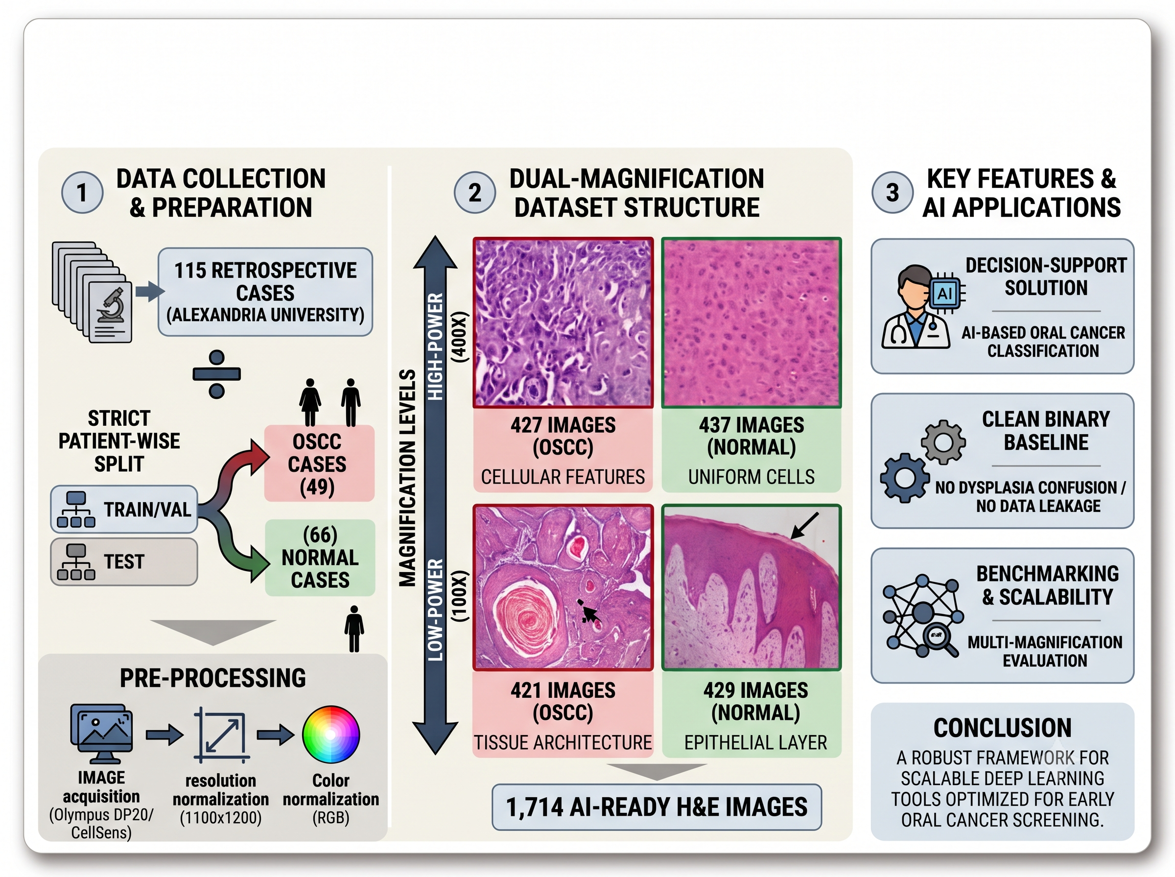
Oral squamous cell carcinoma accounts for nearly 90% of all oral malignancies worldwide. While early detection is critical to improve the disappointed 35%–45% advanced-stage survival rate, manual histopathological evaluation remains a laborious process prone to intra-observer variability and physical strain. Artificial intelligence offers a promising decision-support solution to optimize diagnostic accuracy; however, existing open-source datasets frequently suffer from distinct limitations, including a lack of true normal tissue controls, the confounding grouping of dysplasia with invasive cancer, or a restriction to single-magnification views that fail to mimic a pathologist’s multi-scale workflow. To address these gaps, we introduce a curated, expert-labeled, AI-ready histopathological dataset consisting of 1,714 images obtained from 115 patients, explicitly designed to differentiate between OSCC and completely healthy oral tissue. For clinical usage, the repository provides paired images at low-power magnification to evaluate overall tissue architecture and high-power magnification to distinguish cellular malignancy criteria. Crucially, the dataset is partitioned using a strict patient-wise split rather than an image-level split, successfully eliminating the risk of data leakage during the training, validation, and testing phases of machine learning models. By offering a clean binary baseline with dual-magnification features, this publicly available dataset provides a robust framework for developing scalable deep learning tools optimized for early oral cancer screening. Link of dataset: Bahaa, Salma (2026), “Histopathological images of oral squamous cell carcinoma and normal tissue”, Mendeley Data, V1, doi: 10.17632/7m9zkcx539.1.163
Keywords
Oral Squamous Cell Carcinoma · Histopathology · AI-Ready Dataset
Bibtex
@article{melba:2026:037:bahaa,
    title = "A Histopathological Image Dataset for Oral Squamous Cell Carcinoma and Normal Tissue Analysis",
    author = "Bahaa, Salma",
    journal = "Machine Learning for Biomedical Imaging",
    volume = "2026",
    issue = "Special Issue on MICCAI Open Data 2026",
    year = "2026",
    pages = "758--763",
    issn = "2766-905X",
    doi = "https://doi.org/10.59275/j.melba.2026-273d",
    url = "https://melba-journal.org/2026:037"
}
RIS
TY - JOUR
AU - Bahaa, Salma
PY - 2026
TI - A Histopathological Image Dataset for Oral Squamous Cell Carcinoma and Normal Tissue Analysis
T2 - Machine Learning for Biomedical Imaging
VL - 2026
IS - Special Issue on MICCAI Open Data 2026
SP - 758
EP - 763
SN - 2766-905X
DO - https://doi.org/10.59275/j.melba.2026-273d
UR - https://melba-journal.org/2026:037
ER -