RCC-AID: Renal Cell Carcinoma AI Dataset for Medical Imaging Research
Sarah de Boer1, Hartmut Häntze1,2, Sebastian Ziegelmayer3, Tommaso Russo1,4, Bram van Ginneken1,5, Mathias Prokop1, Keno Bressem3,6, Alessa Hering1
1: Radboudumc, Department of Medical Imaging, Nijmegen, The Netherlands, 2: Charité - Universitätsmedizin Berlin, Department of Radiology, Berlin, Germany, 3: Klinikum rechts der Isar, Department of Diagnostic and interventional Radiology, TUM University Hospital, Technical University of Munich, Munich, Germany, 4: San Raffaele University, Milan, Italy, 5: Fraunhofer MEVIS, Bremen, Germany, 6: German Heart Center, Department of Cardiovascular Radiology and Nuclear Medicine, TUM University Hospital, Technical University of Munich, Munich, Germany
Publication date: 2026/09/21
https://doi.org/10.59275/j.melba.2026-2dg2
Abstract
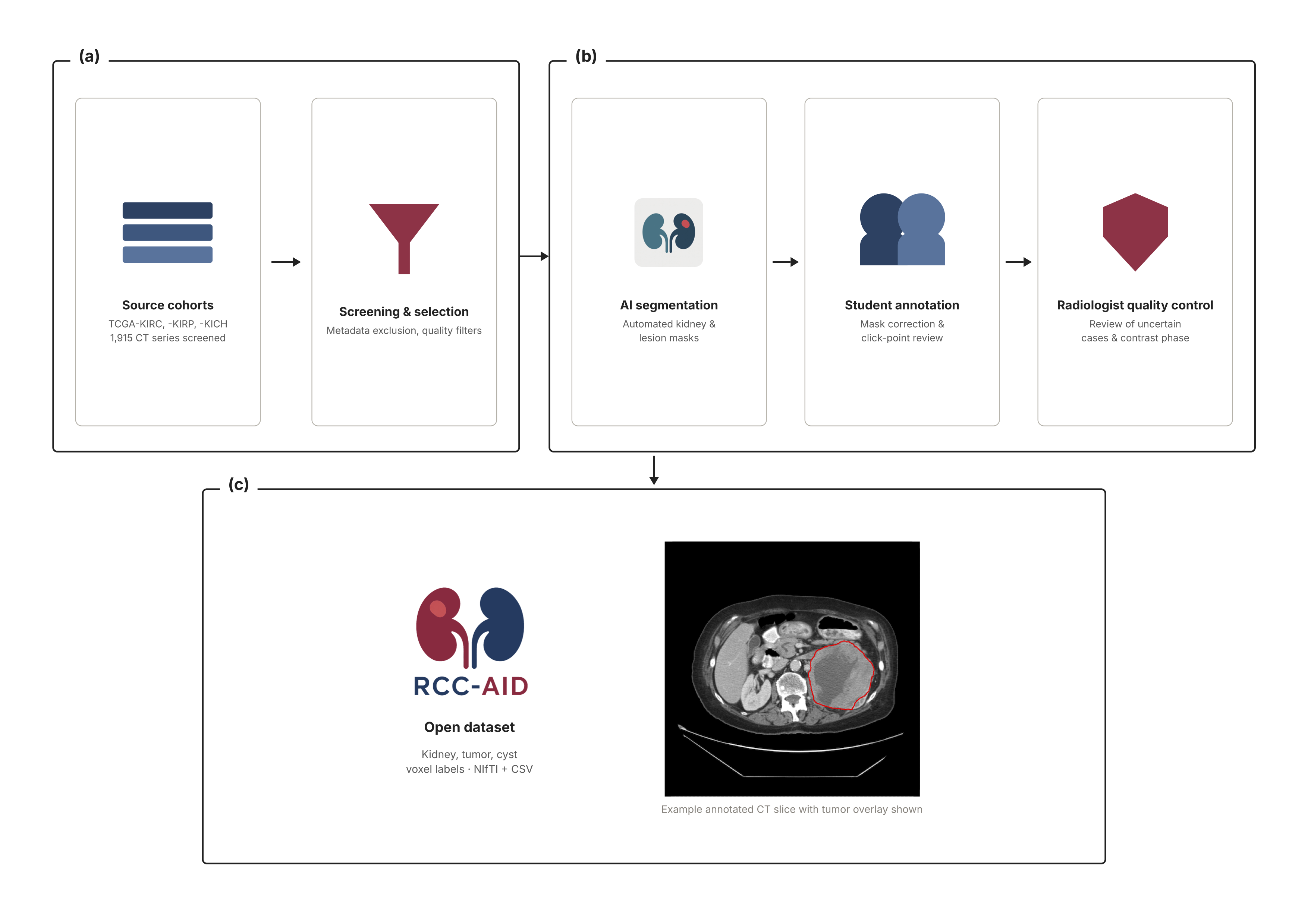
Contrast-enhanced computed tomography (CT) is central to the diagnosis, staging, and follow-up of patients with renal cell carcinoma (RCC). As artificial intelligence research into computer-aided solutions continues to grow, the need for curated and annotated datasets becomes increasingly important. Imaging-based artificial intelligence studies often need lesion annotations that are not consistently available. The Cancer Genome Atlas (TCGA) datasets are widely used for model training and validation. However, access to public annotations of lesions is limited, which limits reproducibility and comparability of the published research. To address this gap, we screened 1,915 CT scans from three TCGA-RCC databases and, following a meta-data-based exclusion step, used an automated segmentation model to generate initial kidney and lesion masks. Next, we conducted a reader study with all papillary (n=56), chromophobe (n=27) and 200 randomly selected clear cell RCC cases. Two trained students performed quality checks, corrections, and additional annotation of tumors and cysts, with uncertain cases reviewed by a board-certified radiologist. After data exclusion and quality control, a final cohort of 129 annotated CT scans from 91 patients (24 female, 67 male; mean age 56 years) was retained, including 85 clear cell, 26 papillary and 18 chromophobe RCC cases. Images and voxel-level annotations of kidneys and lesions are openly available at https://zenodo.org/records/20719257. By open-sourcing these annotations, we aim to foster accessible, reproducible AI research in renal cell carcinoma. RCC-AID provides a reusable open resource dataset for segmentation, detection, subtype classification, radiomics, and multimodal RCC research.
Keywords
Renal Cell Carcinoma · Computed Tomography · Image Segmentation · Open Data · Cancer Imaging · Deep Learning
Bibtex
@article{melba:2026:039:deboer,
    title = "RCC-AID: Renal Cell Carcinoma AI Dataset for Medical Imaging Research",
    author = "de Boer, Sarah and Häntze, Hartmut and Ziegelmayer, Sebastian and Russo, Tommaso and van Ginneken, Bram and Prokop, Mathias and Bressem, Keno and Hering, Alessa",
    journal = "Machine Learning for Biomedical Imaging",
    volume = "2026",
    issue = "Special Issue on MICCAI Open Data 2026",
    year = "2026",
    pages = "772--782",
    issn = "2766-905X",
    doi = "https://doi.org/10.59275/j.melba.2026-2dg2",
    url = "https://melba-journal.org/2026:039"
}
RIS
TY - JOUR
AU - de Boer, Sarah
AU - Häntze, Hartmut
AU - Ziegelmayer, Sebastian
AU - Russo, Tommaso
AU - van Ginneken, Bram
AU - Prokop, Mathias
AU - Bressem, Keno
AU - Hering, Alessa
PY - 2026
TI - RCC-AID: Renal Cell Carcinoma AI Dataset for Medical Imaging Research
T2 - Machine Learning for Biomedical Imaging
VL - 2026
IS - Special Issue on MICCAI Open Data 2026
SP - 772
EP - 782
SN - 2766-905X
DO - https://doi.org/10.59275/j.melba.2026-2dg2
UR - https://melba-journal.org/2026:039
ER -