GLI-AL: A Multi-Modal Glioma MRI Label Resource with Unified Anatomy-Lesion Labels
Xingyu Xiang1,2, Shuang Hao3,2, Fan Wang3,2, Jianhua Ma3,2, Chunfeng Lian1,2
1: School of Mathematics and Statistics, Xi’an Jiaotong University, Xi’an, China, 2: Research Center for Intelligent Medical Equipment and Devices (IMED), Xi’an Jiaotong University, Xi’an 710049, China, 3: Key Laboratory of Biomedical Information Engineering of Ministry of Education, School of Life Science and Technology, Xi’an Jiaotong University, Xi’an, China
Publication date: 2026/09/21
https://doi.org/10.59275/j.melba.2026-575f
Abstract
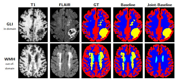
Existing BraTS-GLI datasets provide a widely used benchmark for adult glioma MRI segmentation, but their task definition focuses on tumor subregions and does not systematically represent coexisting white matter hyperintensities (WMH). In joint segmentation settings, such unlabeled abnormalities introduce task-specific label noise by treating pathological regions as normal tissue. To address this limitation, we introduce BraTS-GLI Anatomy-Lesion, a controlled- access, labels-only derived resource built from the BraTS 2023-GLI training cohort. The resource provides 1,251 unified eight-class anatomy-lesion label sets aligned with the original four-modal MRI cases, including image-repair labels for 116 cases requiring repaired imaging inputs. The cohort is organized into a 394-case purified subset and an 857-case extended subset, with case-level metadata covering label source, image-repair requirements, quality-control status, access conditions, checksums, and release boundaries. Compared with the original BraTS-GLI annotations, the resource substantially expands foreground supervision by incorporating healthy brain tissues and previously unlabeled coexisting abnormalities within a unified label space. A validation study using MedNeXt and T1/FLAIR inputs suggests that WMH-aware supervision preserves healthy-tissue segmentation performance across both in-domain GLI and external WMH datasets, while improving sensitivity to coexisting lesions relative to noisy-control training. The resource is intended for scientific research and supports joint anatomy-lesion supervision, label-noise analysis, and reproducible evaluation. Data are available at https://www.synapse.org/Synapse:syn75210889/wiki/, and code is available at https://github.com/xyx200/brats-gli-anatomy-lesion-code. The data resource DOI is https://doi.org/10.7303/SYN75210889.
Keywords
Anatomy-Lesion Segmentation · Label Noise · BraTS-GLI · White Matter Hyperintensities
Bibtex
@article{melba:2026:045:xiang,
    title = "GLI-AL: A Multi-Modal Glioma MRI Label Resource with Unified Anatomy-Lesion Labels",
    author = "Xiang, Xingyu and Hao, Shuang and Wang, Fan and Ma, Jianhua and Lian, Chunfeng",
    journal = "Machine Learning for Biomedical Imaging",
    volume = "2026",
    issue = "Special Issue on MICCAI Open Data 2026",
    year = "2026",
    pages = "834--843",
    issn = "2766-905X",
    doi = "https://doi.org/10.59275/j.melba.2026-575f",
    url = "https://melba-journal.org/2026:045"
}
RIS
TY - JOUR
AU - Xiang, Xingyu
AU - Hao, Shuang
AU - Wang, Fan
AU - Ma, Jianhua
AU - Lian, Chunfeng
PY - 2026
TI - GLI-AL: A Multi-Modal Glioma MRI Label Resource with Unified Anatomy-Lesion Labels
T2 - Machine Learning for Biomedical Imaging
VL - 2026
IS - Special Issue on MICCAI Open Data 2026
SP - 834
EP - 843
SN - 2766-905X
DO - https://doi.org/10.59275/j.melba.2026-575f
UR - https://melba-journal.org/2026:045
ER -